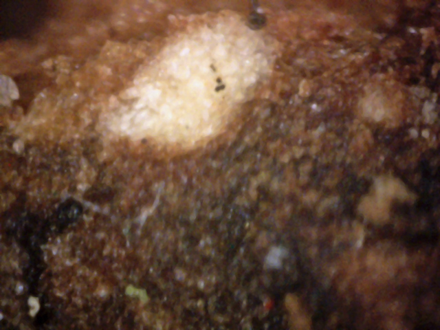
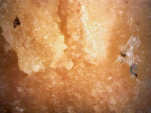
This Red oak sits on the corner of our lot and has been decline for several years.
Being unhappy with several vague diagnoses, I contacted the Cornel plant pathology lab.
They suggested I send in a bark sample. Unsure as to exactly how to do this, and not wanting
to miss any diseased wood, I decided to take a large (6" by 6") and deep sample.
I used a hammer and wood chisel to obtain sample bark around several weep holes.
The following pictures, taken September, 2006, show the sampling process.
1
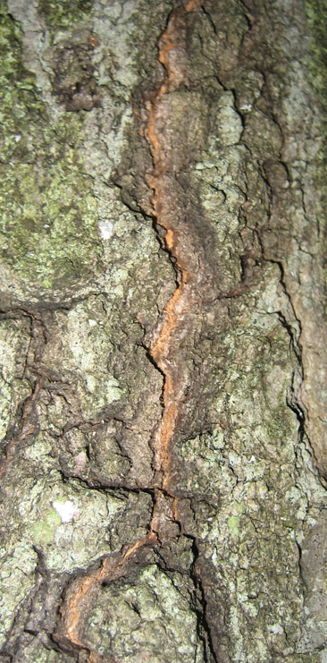
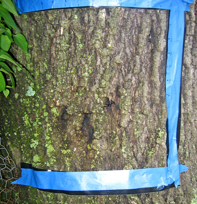
Oaks having White Canker disease often have holes in the bark that they bleed sap from.
Here are three such holes. I decided to obtain a bark sample from this area.
To begin, I marked the area to be sampled with blue tape. Note the inch marks on the tape.
2
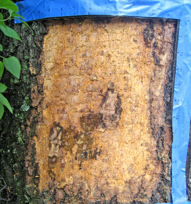
All the outer bark was removed here. The weep spot areas seem to be decayed.
In fact, the decayed area appears much bigger when the outer bark is removed.
3
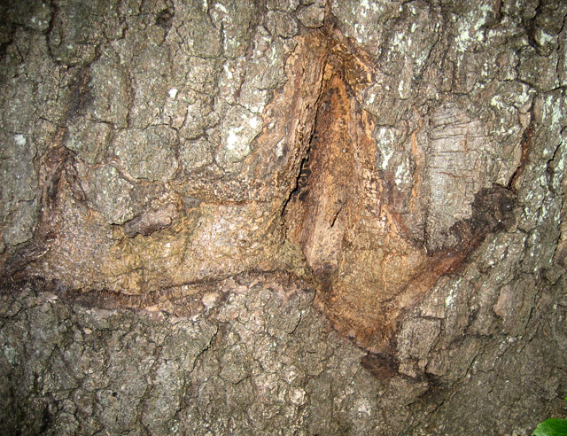
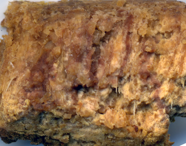
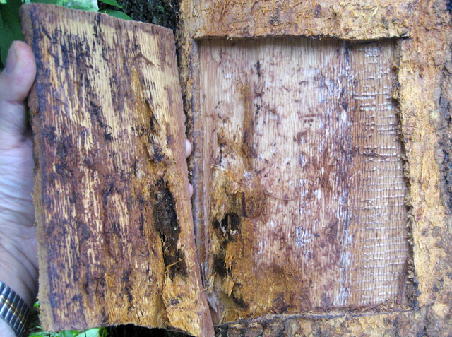
Finally, after much effort, I was able to also remove the inner bark too,
exposing the sapwood. As this picture shows, the disease has burrowed through
all the bark and has continued into the heart of the tree. The wood is black
around the area infected the most.
The Cornell diagnosis was that this tree had a Phytophthora disease (species unknown).
I resumed my investigation in May of 2008, using my scanner to more closely examine parts of the
tree, as shown in the following pictures.

4
This live oak branch is also about 1/4" thick.
The top branch junction was just 6" from the branch tip, with no spores between
it and the tip.
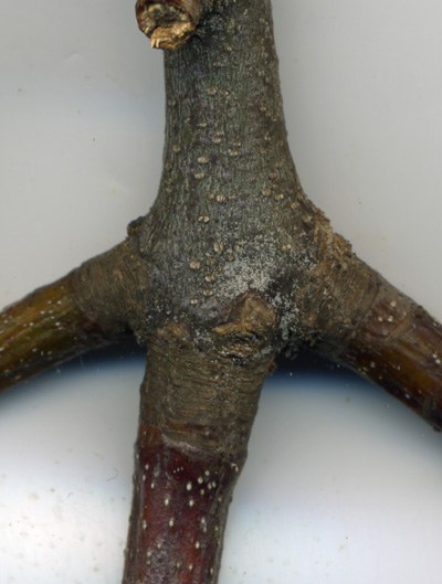
The spores are so thick at the branch junctions and leaf scars that the area appears
almost crusty.
5
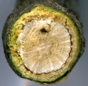
This cross-section of the branch shown in picture 2 was made with a 2400dpi scanner.
The black spot appears to be a disease infection channel.
The spot's location suggests that this tree became infected about 5 years ago (around 2003),
which is about when I started noticing disease symptoms on almost all of my trees and shrubs.

6
This is a dead branch from the red oak tree in decline.
Not only is there a high spore density at the branch junctions and leaf scars,
but the spore density is moderately high between these points too,
making this tree a prime transmitter of disease.
This branch segment was about 1/4" thick, and was located 12" from the branch tip.
There were virtually no spores from this point to the branch tip.
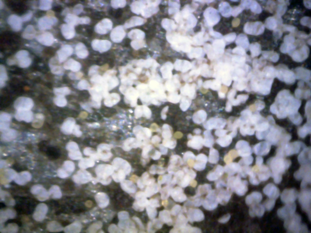
The following set of pictures wase taken in early October 2008, using a 400x digital microscope.
Since I believe the spore release took place shortly after the above pictures were taken, I
didn't really expect to see many spores left.
The first two pictures are of the top of a leaf.
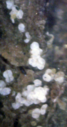
7
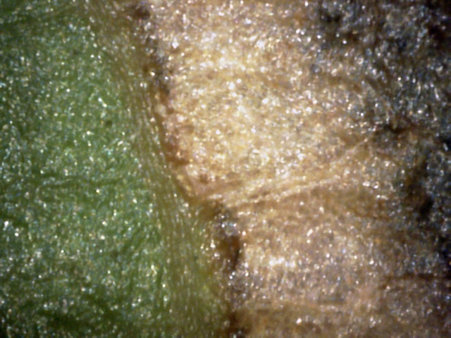
Adjacent live and dead spots on the leaf top.
The dead spot looks like it could be made up of white canker particles.
8
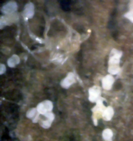
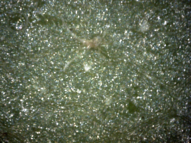
In general, a pretty boring leaf top surface.
As shown here, there were scattered small pieces of white canker, and a few of these
spider-like objects.
Since it's just as easy to check the leaf bottom as the top, the following pictures show
the bottom of the leaf at 400x.
 ←
←
←
←
9
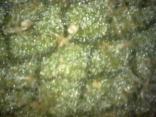
The leaf bottom had numerous spider-like objects scattered about its surface.
But perhaps of even more relevance was the presence of white canker spores (red
arrows), along with their visual characteristics.
The one at center-bottom appears halfway through its embryonic stage, since it's
about half yellow in the center.
The one on top is almost through its embryonic stage since there is only a bit of
yellow showing through its two halves.
 ←
←
←
←
←
←
←
←
←
←
←
←
10
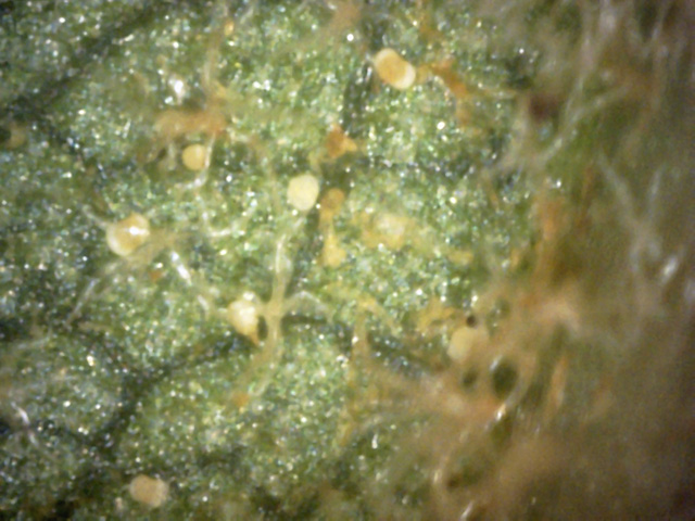
The spores and spider-like objects get more numerous the closer you get to the leaf's stem.
This view is next to the stem.
Of interest here is the variety of development stages of white canker spores.
One (red arrow) is pure yellow and just starting to differentiate.
Three more (blue arrows) are about half-yellow and hence half developed.
Another one (yellow arrow) is almost totally developed, but still has a yellow stalk.
Finally, one (green arrow) is fully developed and totally white.
 ←
←
11
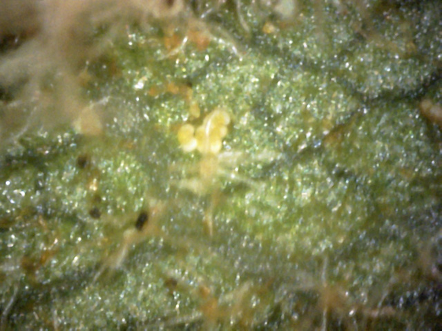
This is another area of the leaf, close to the leaf vein (in the upper-left corner).
Notice the group of developing spores (red arrow) right next to a "spider" object.
Each spore is about half-way through its developing phase, still having a yellow
globular mass within its two halves.
White canker often likes to hide within the body of a leaf. The following set of 6 pictures
check out that possibility via leaf cross-section views at 400x.
 ↓
↓
↓
↑
↓
↓
↓
↑
12
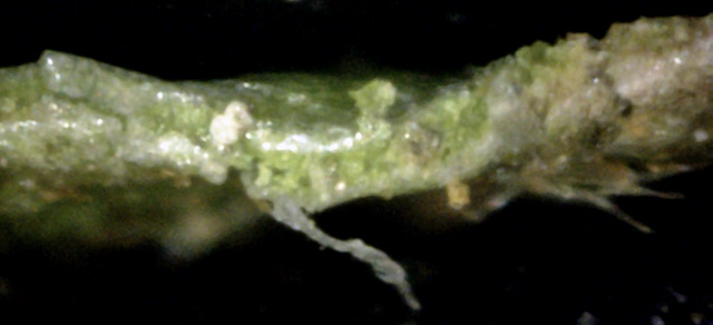
There are two patches of white canker near the center of this picture that run from
the top to the bottom of the leaf (red arrows).
But note the internal gray blob of canker that seems to be giving rise to the spore
on the bottom of the leaf (blue arrow).
There is also a trail of canker (yellow arrow) from this spore to the right and then up to the top
surface of the leaf, where there is another particle of canker material.
 ↑
↓
→
↑
↓
→
13
The left-center part of this leaf's cross-section is of the most interest, as there
is a long "tail" of canker descending from the leaf's bottom (red
arrow).
But where this tail enters the leaf, it gives rise to two spores, one of which appears
to be cut in half (blue arrow).
The stream of canker then continues on left to the top of the leaf.
Again, check the spores - it appears as if one has a white root which intersects
with the gray canker blob.
The intersection forms a tiny yellow spot (yellow arrow).
The question is... did the canker give rise to the spore, or did the spore land on the leaf,
infect it, and then give rise to the canker?
 ←
←
←
←
14
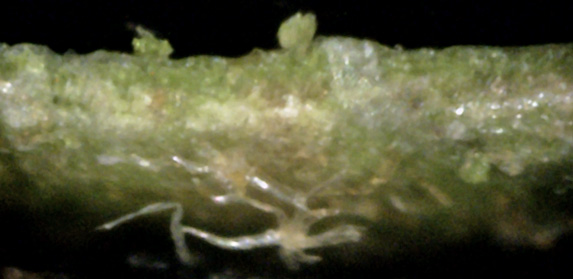
This spider-object appears to have a root (red arrow) going directly into the leaf.
The leaf tissue disturbance hints that the root goes all the way through to the top side,
emitting a green blob (blue arrow) on that surface.
 ↑
↑
15
There are internal blobs of white canker material on the right and left,
that are connected by a tube of white canker (red arrow).
A leaf bottom view wouldn't show this, and a leaf top view
would only show two small disconnected white spots.
This cross-section shows there is a lot of canker within the leaf that isn't visible.
 →
→
16
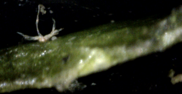
A side view of one of those spider-like objects.
The twisted shape and aspect ratio of the top projection (red arrow) match those of the hyphae
of white canker.
17
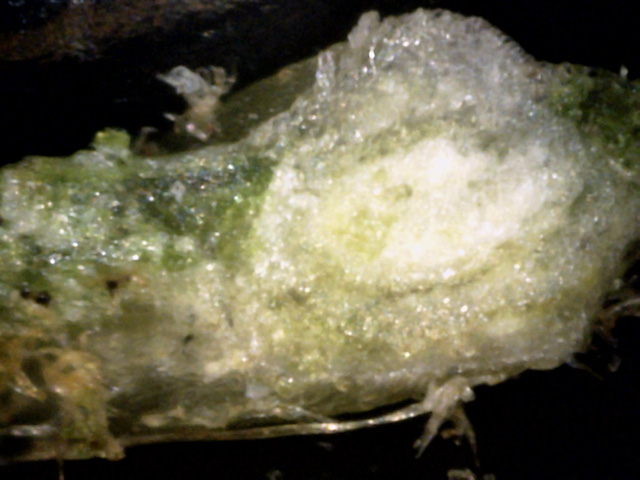
This is a cross-section of the area around a leaf vein.
Going by past experience whereby light-gray-translucent and clear material is canker
material, this leaf vein is about 80% consumed by white canker.
It's not enough to kill the leaf, but it is enough to weaken it severely.
The above set of pictures make a strong case for a white canker infection. But experience has
shown that twig cross-sections provide the best evidence for white canker infections.
The next set of 7 pictures are all cross-section views of a 1/8 inch twig made with a digital microscope
(400x).
 ←
↓
↑
←
↓
↑
18
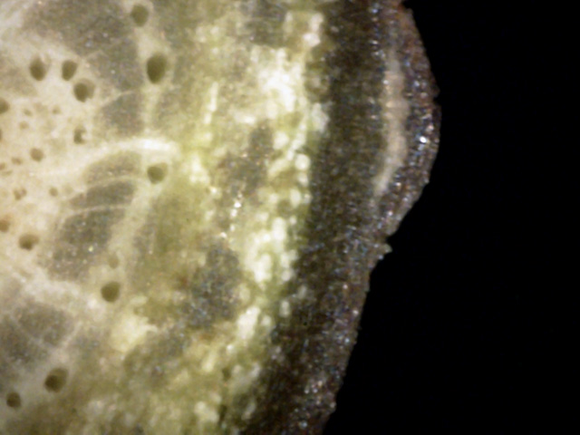
A good solid confirmation of white canker infection.
Particles of white canker have invaded all of the phloem.
A patch of canker has colonized the bark (red arrow), but the tree has grown wound wood around it,
sealing it off.
Two rays appear infected (blue arrows) too, transmitting the infection to the xylem in the center
of the tree.
 ↑
↓
↑
↓
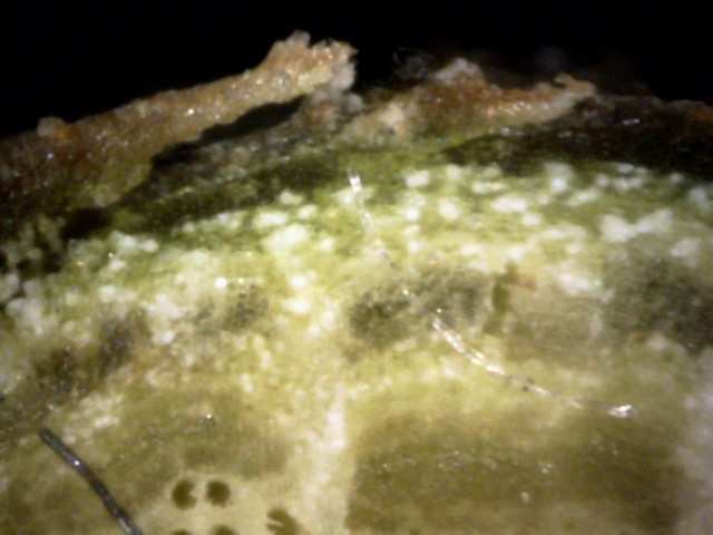
19
Once again, most of the phloem has been infected, and it has been so weakened that the bark
is separating (red arrow) from it. The bark is also infected, so much so that it is turning white
(blue arrow).
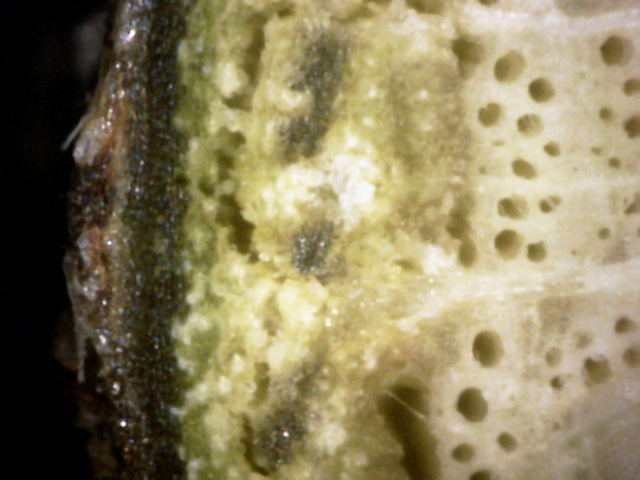
20
White canker has so infected the phloem that it is beginning to shrink and disappear.
The canker is also now spreading into the xylem, destroying it too.
And it has even managed to jump to the outer bark, replacing it with clear white canker.
 ←
←
→
←
←
→
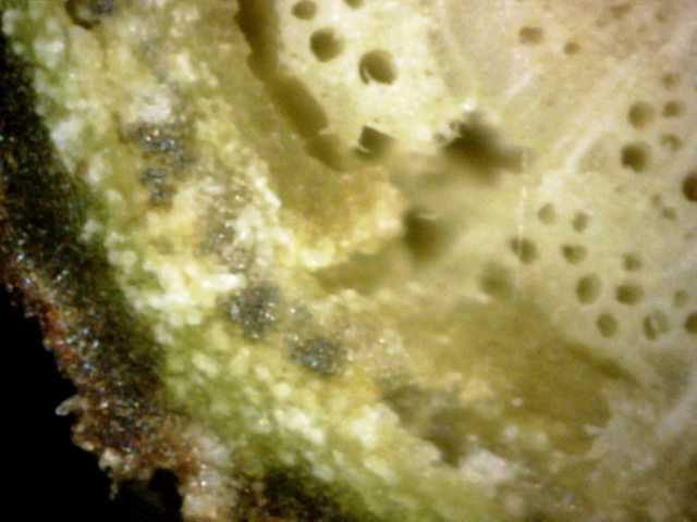
21
Very severe white canker damage is shown here.
The green cortex is heavily infiltrated.
The bark in the lower-left corner has been eaten away and replaced with canker
(red arrow).
The phloem, where it meets the xylem, as turned to a waxy light gray-green color
(blue arrow).
All this canker growth has caused expansion, separating the outer bark and causing
the xylem and phloem to separate (green arrow).
Lacking nourishment, the xylem will no doubt weaken and die.
The branch will then become brittle and easily break off.
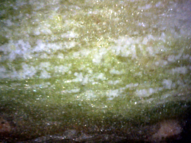
22
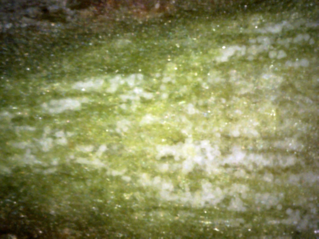
This really isn't a true cross-section, but about a 20 degree slice into the side
of the twig in order to get a better idea of the true shape of this canker.
As seen here, the canker appears as cloud puffs along a string (probably a hypha, actually)
that runs mostly parallel to the twig axis.
Each tiny puff is probably a killed tree cell.
23
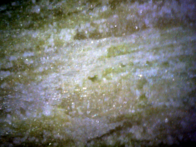
This view is even further along the 20 degree razor cut.
It's clear that the canker much prefers the green phloem layer, but also grows more
weakly and diffusely in the yellow xylem wood.
24
This picture continues further down to the interior of the twig along the 20 degree
(to the twig axis) cross-cut.
Most of the center of the picture is the xylem, and the "puffs of white"
have turned to mostly streaks of white.
That's probably due to the infective hyphae being channeled along the vessels
of the xylem, using them as a sort of "road" to infect other tree tissue.
In summary, while white canker is present in the oak leaves of this tree, the
leaf surfaces were only marginally helpful in diagnosing this white canker
infection since the vast majority of the canker lies within the leaf. Therefore,
a microscopic view of a leaf cross-section more clearly shows the white canker
infection. Nevertheless, the best diagnostic evidence is obtained by examining
the cross-section of a small twig. There the infection is concentrated just
under the bark, at the junction of the light green phloem layer and the dark
green cortex layer. The white canker substance is easily seen there since it
disturbs the natural growth so much.
